The ICTR-PHE 2014 Blog
| Monday | Tuesday | Wednesday | Thursday | Friday |
Day two of ICTR-PHE began with an enlightening talk by Irene Virgolini on the clinical experience with therapies based on the use of radionuclides. Research made in the lab is apt to be implemented in the everyday clinical practice, as underlined by Nicolas Lepareur. The research on new radionuclides and on new types of detectors is crucial in order to obtain clinically applicable techniques, and the first talks of the morning showcased some relevant examples.
Mikael Bourgeois presented the potentiality of the PET emitter 64Cu, a promising radionuclides that can be labelled with a multitude of molecules depending on its use for imaging or pre-therapeutic dosimetry. A very promising application is the one to hypoxia mapping in view of the optimisation of radiation therapy.
Konstantin Zhukov illustrated a gamma probe based on scintillating crystals and silicon photomultipliers that can be used to determine the extent of tumor spread, evaluating which lymph nodes are affected by metastasis, and which are not. Adina Toader presented PIP, the Proton Isotope Producer, an innovative accelerator design based on recirculation, aimed at overcoming the typical issues related to the installation and operation of cyclotrons in the medical environment.
The importance of multidisciplinary collaborations among chemists, biologists, physicists and physicians has been highlighted throughout this session.
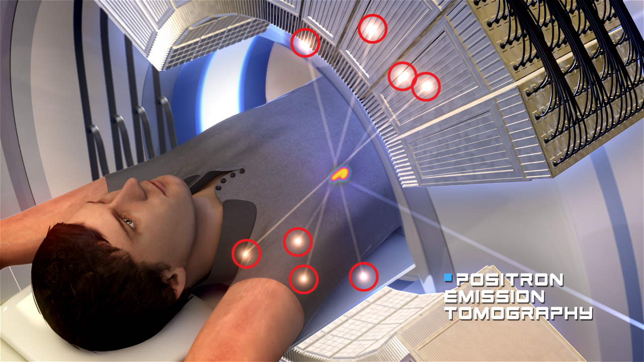
10:30 the session on detectors and imaging begins with an overview talk by David Townsend on multimodality imaging, starting from the first developments on PET and onto the latest developments in PET/MRI.
The second half of the morning focused on imaging and detection, a key element in the development of effective protocols for the treatment of cancer. The session was chaired by Denis Dauvergne and Alberto Del Guerra.
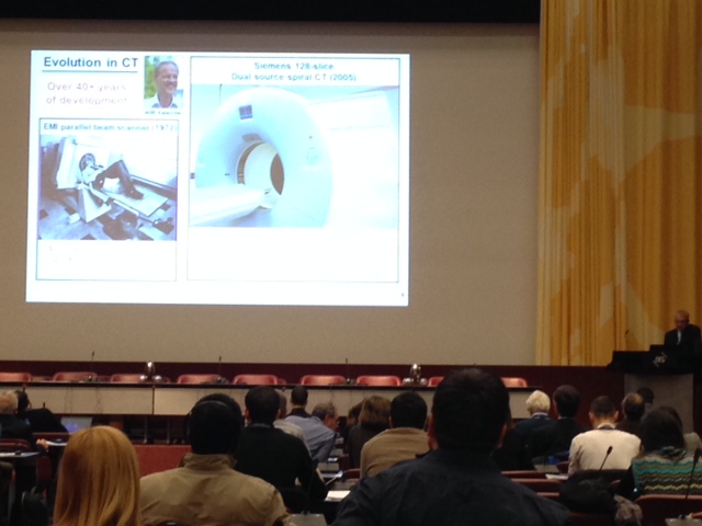
David Townsend offered an inspiring overview of the technologies developed so far, with a specific focus on multimodality imaging. Since PET has been invented in the early 70s, much progress has been made, and the combined use of different technologies is enhancing the quality and the resolution of medical imaging more and more. “Today, 4000 PET/CT Scan are in use worldwide and more under manufacturing,” said Townsend, “and there are more than 6500 publications available PubMed, to document the benefits of this technique.” The next challenge will be the widespread clinical use of PET/MRI. “PET detectors do not like to work in high magnetic fields,” explained Townsend. “But thanks to more than 30 years of research, since 2010 we have PET/MRI machines installed in clinical settings”.
Multimodality imaging is crucial in the development of effective strategies for cancer treatment and screening: in fact, the information that comes from the combination of anatomical and functional imaging data is bigger than the sum of the parts. Beware, though, that we still have to evaluate the real impact of some of these combined techniques: “PET/CT was a technical evolution and an imaging revolution, while PET/MRI represents a technical revolution and an imaging evolution.”
Townsend also highlighted that there are still prejudices related to the amount of dangerous radiation that patients receive with certain imaging techniques, including among radiologists. The overall amount of radiation that patients receive in their lifespan is higher than the single dose received from medical imaging, and several studies proved that chronic exposure can be considered more dangerous than acute delivery of the same dose of radiation.
Besides the technical revolution that made PET/MRI possible, the talk underlined the imperative need to reduce the radiation dose from imaging, not necessarily because it causes cancer, but because people fear that it will cause cancer. This fear has a major impact on healthcare, as patients will refuse to pass some exams, or physicians will prescribe different exams, with less resolution or requiring anesthesia.
Despite the fear, accurate imaging today is considered absolutely safe, and the number of its applications outside the oncology domain is increasing. For instance, “A new drug development can cost up to 5 billion $, but some will not be approved and therefore translated in clinical therapeutic routines. The use of accurate imaging can help to translate new drugs from preclinical to clinical application.”
Imaging has reached spatial resolutions up to 2mm; a further increment in sensitivity will come also from Time of Flight techniques. “We are going toward a very exciting time,” concluded Townsend.
But the quality of the imaging does not only lie in the development of hardware devices: the development of more sophisticated software is crucial in order to integrate the information that comes from the acquisition systems of the different technologies. Dimitris Visvikis explained the challenges and latest developments in this field at large, and showed that morphological data coming from CT or MRI, including 4D acquisition and organs movement and deformation, can be injected in the PET acquisition: the combination of anatomical and functional imaging, using appropriate algorithms, can deliver extremely accurate images.
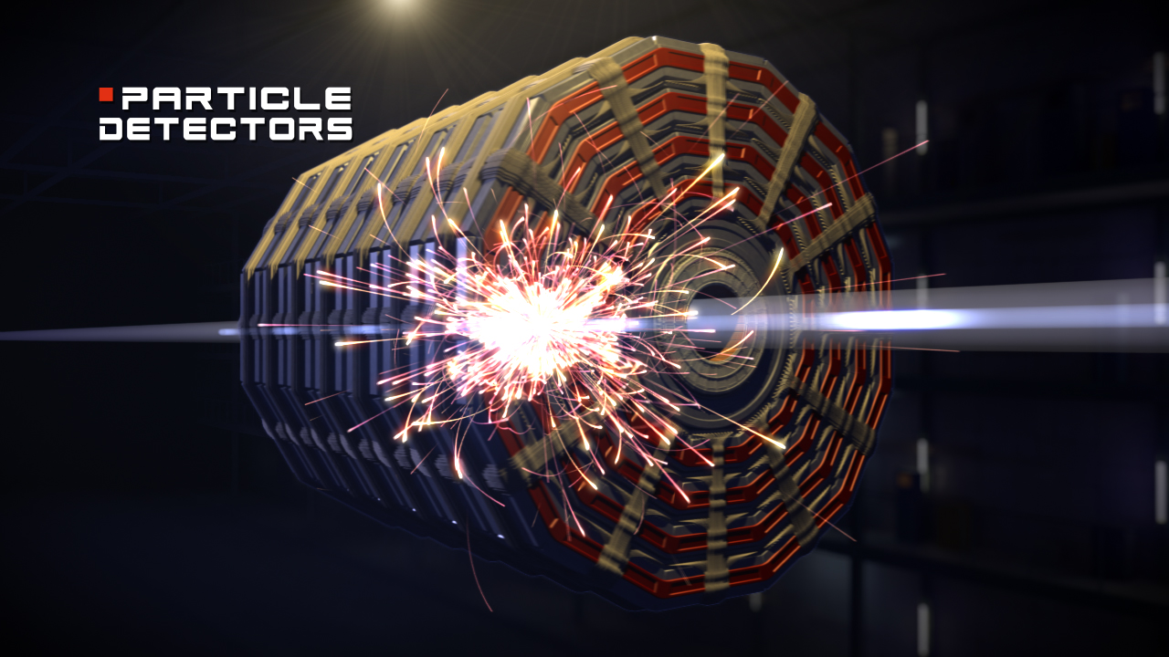
The last talks of the morning focused on beam monitoring, also using innovative approaches. Among these, Piero Giubilato presented a high performance monolithic pixel tracker, able to collect a huge amount of information in a very limited time spread (less than a second to produce a full 3D CT scan) using a compact device assembled starting from commercial technology. “The same technology,” explained Giubilato, “that you can find in your smartphone.”